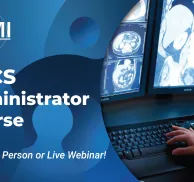

Mammography Registry Review
About this Program
16 CE, Mammography Registry Review 4-part Webinar Series follows the ARRT content specifications as outlined in the most current Mammography Certification Handbook. This webinar series is appropriate for mammographers wanting to prepare for the ARRT Mammography registry or to enhance current knowledge in mammography and breast imaging.
A certificate of attendance, documenting 4-credit hours will be provided for each individual upon completion of each 3.5-hour webinar session. Completing all 4 sessions of this series will provide the 16 hours of structured education related to the content specifications outlined by the ARRT, required for certification and registration.
Session 4 meets 4 hours of the 8-hour MQSA requirement for initial training in digital mammography as it pertains to physicists and radiologic technologists.
A printable download will be available to each registrant prior to each session of the webinar. This will allow you to follow along with the presentation and make notes as you feel necessary.
Tuition listed is per session.
Educational Objectives
At the completion of this webinar series, participants will:
- Understand the differences between regulatory bodies, such as MQSA, ACR and the ARRT.
- Obtain knowledge to help prepare for the ARRT Mammography exam.
- Gain knowledge on the various treatment options for various types of breast diseases.
- Review patient care techniques including documentation of patient history, informed consent and more.
Schedule
In-person and live webinar options
| Session 1 6:30pm Eastern 5:30pm Central 4:30pm Mountain 3:30pm Pacific What's my timezone? | Regulations & Patient Care Accreditation and Certification
Patient Interactions & Management
Evaluation of Image Quality
|
| Session 2 6:30pm Eastern 5:30pm Central 4:30pm Mountain 3:30pm Pacific What's my timezone? | Anatomy & Pathology Localization Terminology
External Anatomy
Internal Anatomy
Cytology
Pathology
|
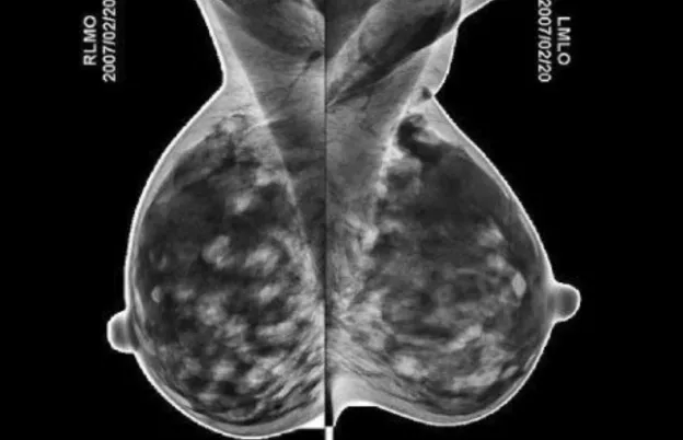
| Session 3 6:30pm Eastern 5:30pm Central 4:30pm Mountain 3:30pm Pacific What's my timezone? | Positioning & Procedures Positioning Views
Special Patient Situations
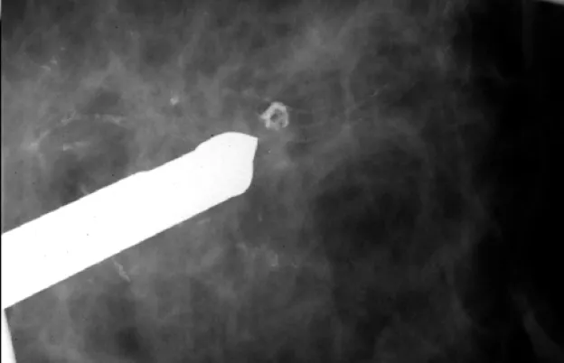
Imaging Examinations
Interventional Procedures
|
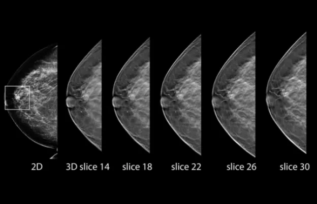
| Session 4 6:30pm Eastern 5:30pm Central 4:30pm Mountain 3:30pm Pacific What's my timezone? | Instrumentation & Quality Control Purpose & Frequency Design Characteristics of Mammography Systems
Mammograhic Technique
Digital Acquisition/Display & Informatics
Mammographer test
|
Audience
Who should attend?
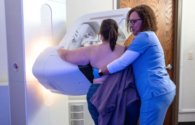
- Mammographers
- Radiologic Technologists
- Managers
- Educators
- Health care professionals involved in providing breast imaging services
- Staff members of companies offering mammographic equipment and supplies
Program Faculty
Meet your presenter(s)

Cheryl Seastrand
RT(R)(M)(BS)
Cheryl Seastrand has over 25 years of experience as a Breast Imaging Specialist. As a registered Mammographer and Breast Sonographer, Cheryl is committed to providing high quality development and delivery of education and training in Breast Imaging. She was on the ARRT Breast Sonography Practice Analysis Advisory Committee and has worked with additional professional affiliations on Mammography projects. Cheryl has a proven history of providing accurate, rational analysis and implementation of regulatory compliance and accreditation. Cheryl works extensively with breast imaging facilities nationally by providing competency assessment and recommended improvements to enhance breast imaging programs.
Though Cheryl Seastrand is an ARRT committee member, by binding contract, Cheryl cannot reveal in whole or in part any ARRT's copyrighted questions or any other insider information about the ARRT's examinations or assessments. The ARRT does not review, evaluate, or endorse review courses, activities, materials, or products and this disclaimer should not be construed as an endorsement by the ARRT.

Miranda M. Lyman-Hager
RT(R)(M)
Miranda became registered with the ARRT as a Radiologic Technologist in 1998 and specialized in Mammography in 1999. She spent the first 8 years working her way up from staff technologist to lead technologist before accepting a Clinic Manager Role with Diagnostic Imaging Centers, P.A. in Lee’s Summit, Missouri in 2006. After 10 years in that role, Miranda took a leap of faith in 2017 and began working as an Independent Mammography Positioning Specialist and Educator. Most of her time was spent teaching for Medical Technology Management Institute (MTMI) on the Initial Mammography Training team as well as traveling the US to provide hands-on positioning workshops. Miranda also worked part-time as Corporate Supervisor in the Mammography Department at University Health, Kansas City, Mo where she collaborated with the Lead Interpreting Physician to ensure all breast imaging protocols, workflows, quality control, and quality assurance issues/concerns were addressed and managed per MQSA/ACR guidelines. In February of 2023, Miranda accepted the position of Director of Women’s Imaging Education at MTMI. In this role, Miranda oversees and manages the Women’s Imaging portfolio of courses and events. Miranda is passionate about women’s health education and continues to provide hands-on positioning workshops as well as teaches several MTMI programs such as Initial Mammography Training, Essential Skills for Quality Mammography, Conquering MQSA and ACR and many more CEU webinars.
Credits
Accredited training programs
ASRT Category A
This program provides 4 hour(s) of Category A continuing education credit for radiologic technologists approved by ASRT and recognized by the ARRT and various licensure states. Category A credit is also recognized for CE credit in Canada. You must attend the entire program to receive your certificate of completion.
A certificate of attendance, documenting 4 hours of credit will be provided for each individual upon completion of each 3.5 hour session of the webinar. Completion of all 4 sessions of this series will provide the 16 hours of structured education related to the content specifications outlined by the ARRT, required for certification and registration.Each session:Biennium CE Credits: 4Structured Education / CQR Credits: 4Tuition
Convenient payment options available
| Audience | Price | Early Price | Member Price | Member Early Price |
|---|---|---|---|---|
| Technologist | $99.00 | $99.00 | $89.00 | $89.00 |
~ Tuition amount listed is per session. ~
Early Pricing Guidelines
Qualifying 'Early' registrations must be made at least 4 days in advance for the program.
Cancellation Policy
Webinars less than 8 hours of credit
Refunds, minus a $15 processing fee, will be granted for cancellations received at least 3 days prior to the program. Cancellations received within 3 days of the webinar will receive a credit toward a future MTMI program, minus the $15 processing fee. No refunds will be made after the webinar starts. MTMI reserves the right to cancel any scheduled program because of low advance registration or other reasons. MTMI’s liability is limited to a refund of any program tuition paid. WEBINAR ATTENDEES that cannot log in due to unsolvable technical issues beyond their control will be eligible for a full refund.
Acknowledgements
Thank you for your support