Bony Pathology: A Technologist Perspective
About this Program
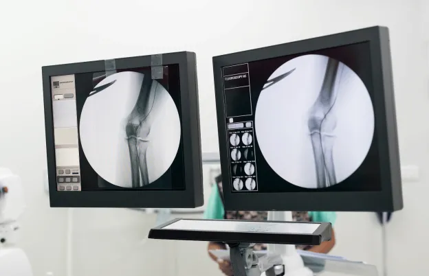
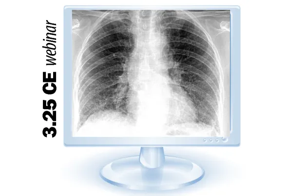
Skeletal radiography still makes up a major portion of medical imaging, principally due to its superior spatial resolution. In this webinar, we analyze bone radiographic images of various body parts using a series of 12 radiographic factors commonly employed by radiologists. Features such as the bony cortex, trabeculae, and periosteum will be studied, as well as bone fractures and neoplasms. A consistent theme in this session will be the role the radiographer plays in optimizing image quality in order to visualize bony pathologies.
Educational Objectives
Upon completion of this session, the participant should be able to:
- Identify the five(5) basic radiographic densities on any radiographic image.
- Explain the interpretive process of radiographic density visualization.
- Discuss low contrast resolution(LCR) and its importance in image interpretation.
- Explain the process of healthy bone homeostasis
- Label microscopic components of an osteocyte.
- Given a radiographic image series, identify important features of healthy bone.
- Analyze bone radiographic images using identified features of bone analysis
- Recognize periosteal reaction, its patterns and its clinical importance
- Distinguish between benign and malignant bone lesions
- Explain the pathological process of bone fracturing and its various types
- Discuss the role of the radiographer in optimizing the diagnostic yield of bone radiographs
Schedule
In-person and live webinar options
Introduction
- Challenges of image interpretation
- Image quality and perceived resolution
- Assessment of bone radiographic image quality
Five Basic Radiographic Densities
- Experiment to demonstrate radiographic densities
- Experiment demonstrating loss of contrast with densities
- Examples of images demonstrating the radiographic densities
Low Contrast Resolution (LCR)
- What is LCR?
- LCR and digital detector technologies
- LCR and image noise
Bone Anatomy and Physiology
- Bone as a tissue
- Cellular structure of bone
- Bone physiology and homeostasis
- Role of periosteum
Radiographic Appearance of Healthy Bone
- Bony cortex
- Bony trabeculae
- Articular surfaces
- Soft tissues of bone
- Periosteum
Twelve Factors of Bone Analysis and Radiographic Image Analysis
- Overall size and shape
- Local size and shape
- Thickness of Cortex
- Trabecular pattern
- General density of whole bone
- Localized density changes
- Margination of local lesions
- Break in cortical continuity
- Periosteal change and appearances
- Soft tissue changes
- Size and contour of joint space
- Alignment of articulating bones
Bony Fractures
- Definition of a fracture
- Importance of views
- Occult fractures and the technologist’s responsibility
- Clinical image analysis of fractures and types
Bone Neoplasms
- Malignant vs. benign
- Radiographic distinctions
- Transition zones and margins
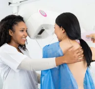
Audience
Who should attend?
- Radiologic Technologists
- Medical Imaging Specialists
- Educators
- Vendor Personnel
Program Faculty
Meet your presenter(s)

Randy Griswold
MPA, RT(R)
Randy has been in the medical imaging profession for over 40 years as an educator, sales and marketing professional and consultant. Currently he is a contributing author and medical imaging consultant. Prior to that, he was the Director of Sales and Marketing for a Midwest distributor of digital medical imaging products. His collective experiences as an educator include being the Program Director for Bellin College, School of Radiologic Sciences and their BSRS program. He was instrumental in transforming their two-year certificate program to a four-year, accredited BSRS degree program.
Randy has 23 years of experience in radiology capital equipment sales, service and support including digital imaging, and has completed his graduate work in Public Service Administration with an emphasis in Health Care Administration and Medical Imaging Marketing. He is a past president of the Wisconsin Association of Educators in Radiologic Technology (WAERT) and the Wisconsin Society of Radiologic Technologists (WSRT) as well as being a Fellow. His passion for teaching is focused on helping technologists understand the importance of obtaining good quality images for diagnosis, in a fashion that uses the skills and techniques of radiologist interpretation.
Credits
Accredited training programs

ASRT Category A
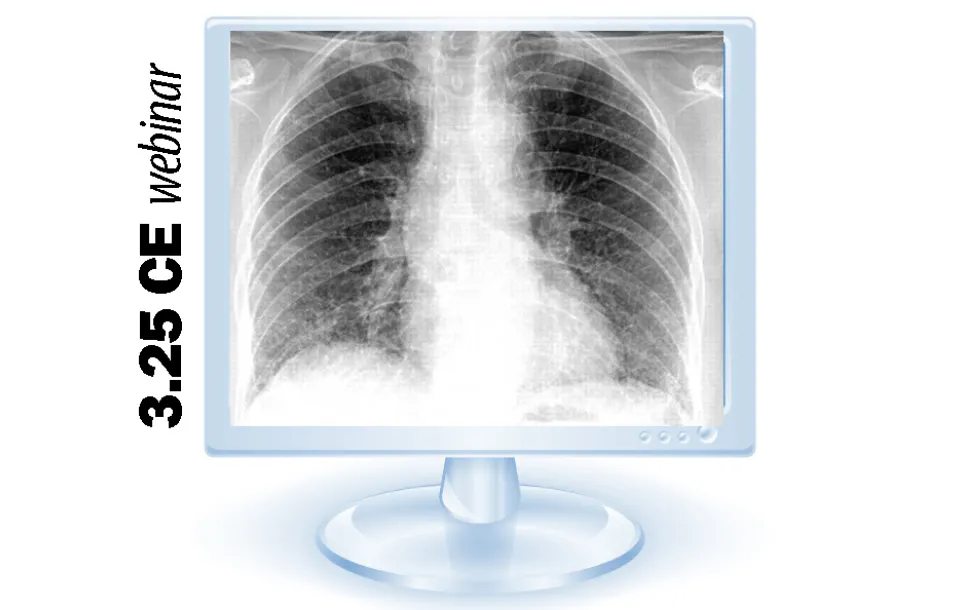
This program provides 3.25 hour(s) of Category A continuing education credit for radiologic technologists approved by ASRT and recognized by the ARRT and various licensure states. Category A credit is also recognized for CE credit in Canada. You must attend the entire program to receive your certificate of completion.
Tuition
Convenient payment options available
| Audience | Price | Early Price | Member Price | Member Early Price |
|---|---|---|---|---|
| Technologist | $69.00 | $65.00 | $62.00 | $59.00 |
Early Pricing Guidelines
Qualifying 'Early' registrations must be made at least 4 days in advance for the program.Cancellation Policy
Webinars less than 8 hours of credit
Refunds, minus a $15 processing fee, will be granted for cancellations received at least 3 days prior to the program. Cancellations received within 3 days of the webinar will receive a credit toward a future MTMI program, minus the $15 processing fee. No refunds will be made after the webinar starts. MTMI reserves the right to cancel any scheduled program because of low advance registration or other reasons. MTMI’s liability is limited to a refund of any program tuition paid. WEBINAR ATTENDEES that cannot log in due to unsolvable technical issues beyond their control will be eligible for a full refund.