
Most people, at least in the U.S., have had at least one medical imaging study in their lifetime, a clear indicator of the impact radiology has had on public health. In 2015, this science celebrated its 100th birthday. That makes it sound old, but the truth is that radiology is still relatively new and evolving more and more each passing year as desirable medical outcomes continue to improve.
It started in the late 1800s and grew into the digital revolution we see today. If you are looking for a career in healthcare that puts you on the cutting edge, then radiology might be for you. How did x-ray get its start?
What Is Radiology?
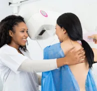
Radiology is a medical specialty that creates and interprets pictures of the human body's organs and body systems to diagnose sickness or injury. Radiology, as it is commonly called, also can be referred to as imaging services, medical imaging or diagnostic imaging. The common term here is 'imaging' since that is its focus. It may consist of x-ray technologies such as radiography, CT scanning, DXA scanning, mammography, angiography and interventional medicine. Additionally, non-x-ray technologies such as ultrasound, radionuclide imaging, and magnetic resonance (MR) scanning also make up the radiology family.
In many ways, radiology serves every healthcare sector, including emergency medicine, pediatrics, trauma response, infectious disease, orthopedics, dentistry, orthodontics, obstetrics, and cancer care, oftentimes referred to as oncology.
The field is divided into diagnostic radiology and therapeutic radiology, both of which employ radiation to detect and cure disorders. While there are several imaging tests available, some of the most popular include x-rays, MRIs, ultrasounds, CT scans, and PET scans.
History of Radiology Timeline
Even with just over 100 years of science under its belt, radiology has seen some fascinating discoveries beginning with the X-ray followed by fluoroscopy, computed radiography, and digital radiography.
Who Discovered the X-Ray?
The story of radiology begins with Wilhelm Conrad Roentgen, a German mechanical engineer and physicist. Roentgen discovered a new type of electromagnetic energy as he was experimenting in his laboratory. It was a discovery for which he would later win a Nobel Prize in Physics in 1901.
On November 8, 1895, Roentgen was studying the behavior of electrons as they passed through a glass tube, known as a Crookes-Hittorf tube. The tube was completely enclosed in a light-tight cardboard wrap and his work was performed in near total darkness in his laboratory. As he experimented with the tube, he accidentally noticed a nearby workbench coated with barium platinocyanide, giving off a low intensity, greenish light. The light seemed to happen as the electron tube was energized and he quickly made the association between the flow of electrons and this “New Kind of Ray.”
During further tests, he found that objects of varying thickness placed in the path of the rays revealed changing transparency when captured on a photographic plate. When he put his wife's hand in the path of the rays over a photographic plate for a few moments, it developed a unique image.
The developed plate showed shadows thrown by the bones of her hand and a ring she was wearing, surrounded by flesh, which was more permeable to the rays and thus threw a fainter shadow. That was the first X-ray image of a human being. Over the next several days, Roengten worked to near exhaustion and discovered most all of the properties of x-rays, that still are true to this day. He called them “x-rays” with a plan to name them more accurately at a later time, but that never happened. His work and dedication are a testament to the brilliance of Wilhelm Roengten as a physicist and his quest for scientific answers.
Several scientists, including Thomas Edison, have built on the inventions of Wilhelm Roentgen. Edison created fluoroscopy around the turn of the century. Fluoroscopy uses X-rays to create moving images in real-time. Unfortunately, the dangerous effects of intense, long-term x-ray exposure became evident with the death of Edison’s assistant, Clarence Dally. This paved the way ultimately to a better understanding of the harmful effects of x-rays and proper radiation safety. In 1918, George Eastman introduced film to replace the glass photographic plates used in an X-ray. Eventually, these impractical glass plates were replaced with highly-flammable, cellulose nitrate sheets that finally were replaced with stable polyester films. Nowadays, x-ray images are digital in nature and the polyester film has become obsolete. Radiology is truly a “digital imaging technology.”
Ultrasound Technology
Ultrasound, often referred to as medical sonography, is the use of ultra-high frequency sound waves for medical procedures. These sound frequencies are well above the human audible range and safe for human applications. Although Scottish obstetrician Ian Donald created the first ultrasound in 1958, the technology goes back to Jacques and Pierre Curie in 1877. They were the first to use piezoelectricity, converting kinetic or mechanical energy into electrical energy. This is a critical component of ultrasound transducers.
In 1958, Dr. Ian Donald used this technology to observe the growth of fetuses in the womb to visualize abnormalities during pregnancy. Donald’s interest in ultrasound technology began when he met the director of a boiler fabrication company.
The company used an industrial ultrasound to check for cracks in their welds. Donald was curious to know if the ultrasound could differentiate between different types of tissue. He visited the plant in July of 1955, bringing with him fibroids and a large ovarian cyst. He used the ultrasound machine on these tissue samples, comparing them with a steak as a control.
The experiment confirmed the machine’s ability to scan biological tissue. Along with other contributors, Donald built a smaller version of the ultrasound to use on women expecting babies. The modern-day ultrasound expands beyond obstetrics, though, and can effectively visualize most parts of the body, including the heart, blood vessels and abdomen. Most recently, higher sound frequencies are being used for musculoskeletal (MSK) studies which are clinically important to orthopedic physicians.
Tomography
In the 1930s, radiologist Dr. Alessandro Vallebona, director of the Institute of Radiology, presented a method for anatomically representing a single body slice on radiographic film. Tomography is the use of x-rays to create images. The word comes from the Greek words "tomos," which means "slice" or "section," and "graphia," which means "describing." The creation of x-ray image data sets as slices, overcomes the superimposition of human anatomy on images, which can be confusing to radiologists.
Today, tomography is a science used by everyone from archaeologists to oceanographers and has wide applications in diagnostic medicine. Scientists have built upon Dr. Vallebona’s work further to create computed tomography.
Computed Tomography
Computed tomography is what most people know as a CT or CAT scan. CTs use the same technology as X-rays but produce images in a cross-section. Created in 1972 by Oxford mathematicians Sir Godfrey Hounsfield and Allan Cormack, CT scans can detect conditions not seen in a conventional X-ray, such as:
- Structures in the brain including tumors
- Blood clots
- Enlarged ventricles
- Abnormalities in the nerves or muscles
- Abdominal masses
- Bone fractures
The CT scanner focuses a narrow x-ray beam through the patient, as a sophisticated x-ray detector and x-ray tube assembly, rotates around the patient in sub-second times. This creates CT data sets of human anatomy on a slice-by-slice basis, analogous to “slicing a loaf of bread.” Different densities of the various structures and organs absorb the x-rays in varying degrees, a process known as differential absorption, creating images displayed on computer screens for interpretation. Many times, these images can be displayed in 3-D.
In 1979, Sir Hounsfield and Cormack were awarded the Nobel Prize in Physiology or Medicine for their contributions to health and research.
Magnetic Resonance Imaging
Dr. Raymond Vahan Damadian is an American physician and the developer of the first MRI scanning machine. He first proposed the idea of the MRI in 1969. MR scanning, as it is commonly called, relies upon the atomic characteristics of magnetism and radiofrequencies to create images at the atomic level, of cells and tissues that contain water, and in particular hydrogen, as an essential element of water. MR scanners look similar to CT scanners but the physical principles of operation are quite different. MR technology does not use radiation and is able to reveal the state of tissues and disease at the cellular level, by using various MR sequences.
Dr. Damadian showed that malignancies and normal tissue might be discriminated in vivo by nuclear magnetic resonance (NMR) because of their longer atomic relaxation durations. He performed the first full-body MRI on a human in 1977 to identify cancer. MR scanning has revolutionized medicine from a diagnostic point and is now an essential tool in medicine.
Digital and Computed Radiology
These incredible imaging technologies have led to even further advances such as digital (DR) and computed (CR) radiography. Both necessitate the use of digital technologies that rely on computer networks and high-bandwidth internet connections.
DR employs flat panel detectors and thin film transistor technology that convert X-rays to electrical charges directly or indirectly, which are subsequently computer processed using complex software programs to generate a digital picture using x-rays. Space-age materials such as selenium, silicon and perovskite are finding applications with DR detectors as the industry continues to advance and search for higher image quality and lower radiation dosages.
CR uses cassette-based phosphor storage plates (PSP) which are then scanned into a digital format by a computerized system for picture processing, archiving, and display. With both CR and DR, the entire operation is digitized to produce radiographic images that can be networked and transmitted world-wide for consultation and interpretation. As digital data sets, these images are archived for long-term storage, in sophisticated picture archival and communication systems (PACS).
Positron Emission Tomography (PET)
A PET scan is a type of imaging that uses radionuclides to reveal the metabolic or biochemical activity of tissues and organs. A radioactive substance (tracer) such as Fluorine-18 is used in the PET scan to show normal and abnormal metabolic activity. PET scanning relies upon the atomic reaction of pair-production to create images using a nuclear “gamma detector.” A PET-CT scan combines a CT with a PET scanner and is generally more accurate in diagnosing cancer. PET is often used to help stage cancers and assist oncologists in developing chemo and radio-therapy treatment plans.
Future of Radiology
The technology involved in radiology is transformative and the pace of innovation continues to accelerate. You can expect to see artificial intelligence (AI) play a significant role in this science in the future.
AI will be integrated into radiologists' regular practices, assisting physicians in improving efficiency and diagnostic capabilities. AI can filter through massive amounts of imaging data in seconds, supporting radiologists by prioritizing worklists and diagnoses.
Also in the works is magnetic resonance microscopy (MRM) for deep tissue imaging. MRM is unique in providing cellular-level information from tissue many centimeters deep. The utilization of specialized hardware, ultra-high fields, and novel data collecting and processing algorithms creates a one-of-a-kind method for extracting biophysical information at the cellular level in intact samples.
Another exciting offshoot is medical imaging informatics. This branch of biomedical informatics collects, analyzes, and communicates essential imaging information critical to patient care.
For more than 100 years, radiology has combined the best of technology and science to improve research and patient care. To find out more about careers in this exciting field, check out the programs available at the Medical Technology Management Institute (MTMI). You can contact us today to find out more.