Breast Ultrasound Workshop
For more information or to schedule your in-service program,
Call (800)765-6864 or email custservice@mtmi.net
About this Program
Information on this page is an example of the Breast Ultrasound program that MTMI can provide. A customized program can be created to meet the needs of your facility.
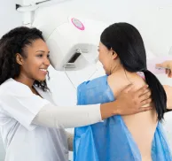
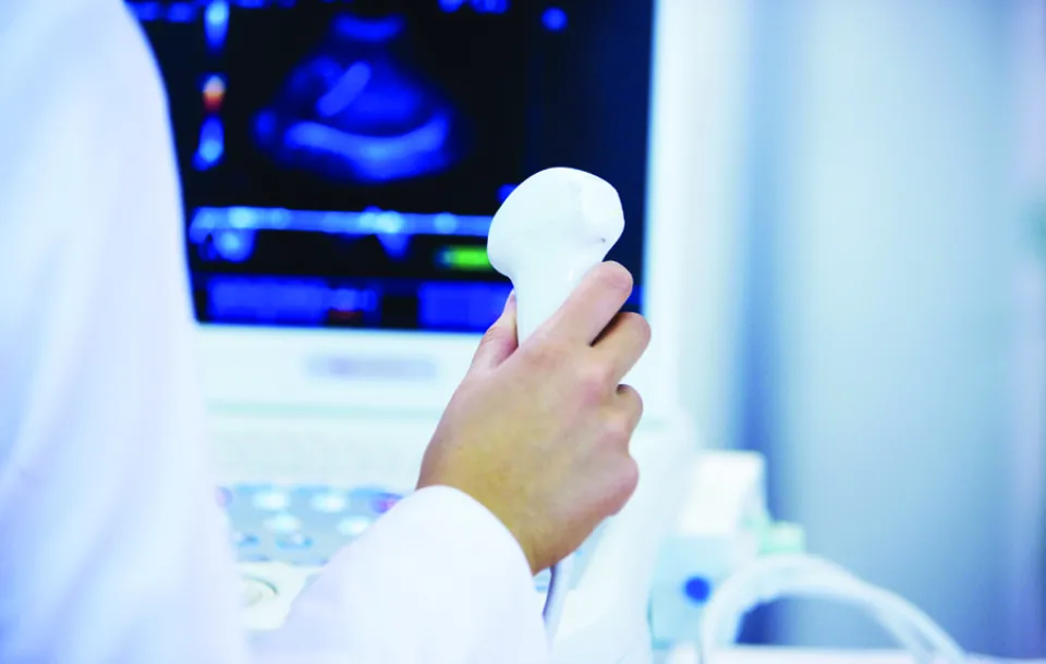
This course will provide increased knowledge of the technical and clinical aspects of breast ultrasound as well as practical hands-on training of ultrasound imaging of the breast. An overview of the basic principles and techniques utilized in breast ultrasound will be presented. This course will be helpful for mammographers and sonographers who are directly involved in breast imaging and physicians whose practice includes women’s health.
Institutional Requirements
A meeting room or class room
Promotion of the event to internal staff
An on-site meeting coordinator
Audio-visual equipment
- Laser pointer
- Screen
- Microphone
- Laser pointer
- Screen
- Microphone
Educational Objectives
- Understand how the basic principles and instrumentation of ultrasound affect the technique of breast ultrasound.
- Recognize normal breast anatomy on the ultrasound image and evaluate lesions for their benign or malignant features.
- Recall the mammographic and ultrasonic features of specific benign and malignant lesions.
- Explain the role and importance of mammography, ultrasound, MRI, nuclear medicine, and ductography in the work up and management of the breast.
- Recognize breast ultrasound artifacts, understand why they occur, and describe techniques to minimize the occurrence of those that degrade the image.
- Develop an awareness of the invasive procedures that are available, as well as, the benefits and limitations of each.
- Define the different surgical and nonsurgical treatment options for patients who develop breast cancer.
Schedule
What this course will cover
| SAMPLE SCHEDULE | |||||||||||||||||||||||||||||||||||||||||||||
| Day One | Day Two | ||||||||||||||||||||||||||||||||||||||||||||
|
|
Program Faculty
Meet your presenter(s)

Cheryl Seastrand
R.T.(R)(M)(BS)(ARRT)
Cheryl is a registered mammographer and breast sonographer with the ARRT and has worked in medical imaging for 39 years. She was a lead Breast Imaging Technologist at Aurora St. Luke’s Medical Center in Milwaukee, WI and was also a charter member of Aurora Mammography Workshops planning team. Cheryl has worked with MTMI on the breast ultrasound initial training course from 2013-2024, as well as developing various breast ultrasound on-site in-services to assist with image optimization for technologists and physicians. She has also worked with MTMI on the mammography initial training and registry review courses, provided mammography accreditation in-services, as well as developing variety of videos and webinars during the past 12 years. Cheryl is married and has one daughter who is married and lives in Indianapolis, IN.
Credits
Accredited training programs
This course provides 16 hours of Category A continuing education credit for radiologic technologists and sonographers. Category A credit from the ASRT meets the requirements for both the ARRT and the ARDMS. A certificate of attendance will be provided for each individual upon completion of the program. All attendees must attend the entire program to receive credit. Credit from various licensure states has been approved.
The Medical Technology Management Institute is accredited by the Accreditation Council for Continuing Medical Education to provide continuing medical education for physicians.
The Medical Technology Management Institute designates this live activity for a maximum of 16 AMA PRA Category 1 Credits ™. Physicians should claim only the credit commensurate with the extent of their participation in the activity.