Basic Mammographic Positioning- Craniocaudal & Mediolateral Oblique Views- Solis
Credits
- 1.5 ASRT Category A
About this Program
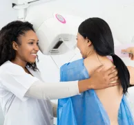
This program we will be discussing and demonstrating mammographic positioning techniques for the craniocaudal and mediolateral oblique views. It is the goal and the responsibility of mammographers to strive to include as much breast tissue as possible while demonstrating the correct anatomy and pathology. This program will provide you with the proper tools to enhance your current positioning
Agenda
Overview of Mammographic Positioning
- Identify the two standard views for mammography - Optimal Craniocaudal (CC) and Mediolateral Oblique (MLO).
- Describe the standard projections for CC and MLO.
- Identify the limitations of the standard projections for CC and MLO.
- Define the mobility principle in mammographic positioning.
- Describe mammographic compression and its benefits.
Basic Mammographic Positioning – Craniocaudal (CC)
- describe the mammographic positioning and compression techniques for Craniocaudal Oblique view.
Basic Mammographic Positioning – Mediolateral Oblique (MLO)
- describe the mammographic positioning and compression techniques for Mediolateral Oblique view.
“This CE activity may be available in multiple formats or from different CE sponsors. ARRT does not allow self-learning CE activities (e.g., Internet courses, home study programs, directed readings) to be repeated for CE credit in the same CE biennium.”
How it Works:
- Your On Demand CE activity that you purchased will be located in your “My Account” section once you log into the MTMI Website.
- You have three attempts to pass each quiz.
- You must earn a score of 75 % or higher.
- Credit is recorded the day you submit and pass the quiz and is determined using Central time.
- You have 30 days to complete and pass the quiz.
- Once passed, access your MTMI “My Account” to print your “Certificate of Completion.”
Educational Objectives
By the end of this program, the participant should be able to:
- Demonstrate proper positioning techniques for the craniocaudal and mediolateral oblique views in mammography.
- Identify techniques that can be used to optimally visualize all posterior glandular tissue and retroglandular fat on the craniocaudal view.
- Identify techniques that can be used to optimally visualize the posterior tissue, retroglandular fat and pectoral muscle on the mediolateral oblique views.
Program Faculty
Meet your presenter(s)

Cheryl Seastrand
R.T.(R)(M)(BS)(ARRT)
Cheryl is a registered mammographer and breast sonographer with the ARRT and has worked in medical imaging for 39 years. She was a lead Breast Imaging Technologist at Aurora St. Luke’s Medical Center in Milwaukee, WI and was also a charter member of Aurora Mammography Workshops planning team. Cheryl has worked with MTMI on the breast ultrasound initial training course from 2013-2024, as well as developing various breast ultrasound on-site in-services to assist with image optimization for technologists and physicians. She has also worked with MTMI on the mammography initial training and registry review courses, provided mammography accreditation in-services, as well as developing variety of videos and webinars during the past 12 years. Cheryl is married and has one daughter who is married and lives in Indianapolis, IN.
Credits
Accredited training programs
ASRT Category A
This program provides 1.5 hour(s) of Category A continuing education credit for radiologic technologists approved by ASRT and recognized by the ARRT® and various licensure states. Category A credit is also recognized for CE credit in Canada. You must attend the entire program to receive your certificate of completion.
