

From Densities to Diagnosis: The Hidden Insights of Radiography - OnDemand
Credits
- 2.0 ASRT Category A
About this Program
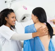
Radiographic images are more than black and white—they’re layered with diagnostic clues that can be easily missed without a deeper understanding of tissue densities and contrast. In this advanced-level course, Randy Griswold, MPA, RT(R), leads participants on a comprehensive exploration of the five basic radiographic densities and the critical role of adipose tissue in image interpretation.
Through engaging demonstrations and real clinical examples, learners will enhance their understanding of image quality, low contrast resolution (LCR), and the subtle but diagnostic significance of fat pads and fat stripes across various anatomical regions. From recognizing early signs of injury, such as lipohemoarthrosis, to identifying anatomical asymmetries, this course equips radiologic technologists with tools to refine their diagnostic awareness and interpretive assessment skills.
Agenda:
- Introduction
- Challenges of Image Interpretation
- Image Quality and Perceived Resolution
- Assessment of Image Quality
- Five Basic Radiographic Densities
- Experiment to Demonstrate Radiographic Densities
- Experiment Demonstrating Loss of Contrast with Densities
- Examples of Images Demonstrating the Radiographic Densities
- Adipose as a Tissue Density
- Average Atomic Density (Z#)
- Comparison to Other Tissue Densities and Z#
- Low Contrast Resolution (LCR)
- What is LCR?
- LCR and Digital Detector Technologies
- LCR and Image Noise
- Images of Experiment on LCR and Beam Hardening
- Clinical Significance of Fat
- Elbow Fat Pads
- Paraspinal Fat Stripes
- Renal Fat and Fascia
- Properitoneal Fat Stripes
- Hoffa’s Fat Pad
- Kager’s Triangle
- Pronator Fat Stripe
- Clinical Examples of Radiographic Fat
- Lipohemoarthrosis
- Explanation and Clinical Significance
- Importance of Positioning and Patient at Rest
- Ship’s Sail Sign
- Importance of Positioning
- Asymmetry of Fat Stripes
- Effusion of Fat Compartments and Radiographic Visualization
- Lipohemoarthrosis
How it Works:
- The On Demand CE activity that you purchased will be located in your “My Account” section once you log into the MTMI Website.
- You have three attempts to pass each quiz.
- You must earn a score of 75% or higher.
- Credit is recorded the day you submit and pass the quiz and is determined using Central time.
- You have 30 days to complete and pass the quiz.
- Once passed, access your MTMI “My Account” to print your “Certificate of Completion.”
- This video expires 1 year after purchase date.
Educational Objectives
At the conclusion of this program, the participant should be able to:
- Identify the five (5) basic radiographic densities on any radiographic image
- Explain the interpretive process of radiographic density visualization
- Discuss low contrast resolution (LCR) and its importance in image interpretation
- Explain the clinical importance of adipose in various body compartments, from a diagnostic viewpoint
- Identify several body structures that demonstrate radiographic shadow formation based upon adipose distribution
- Explain the radiographic clinical interpretive importance of adipose in the elbow, knee, ankle, and wrist joints, as well as the abdomen
- Discuss the development of lipohemoarthrosis and its radiographic and clinical significance
- Point out the process of tissue effusion and its impact on fat visualization
- Suggest methods/techniques that could be utilized to optimize LCR and adipose visualization
Program Faculty
Meet your presenter(s)

Randy Griswold
MPA, RT(R)
Randy has been in the medical imaging profession for over 40 years as an educator, sales and marketing professional and consultant. Currently he is a contributing author and medical imaging consultant. Prior to that, he was the Director of Sales and Marketing for a Midwest distributor of digital medical imaging products. His collective experiences as an educator include being the Program Director for Bellin College, School of Radiologic Sciences and their BSRS program. He was instrumental in transforming their two-year certificate program to a four-year, accredited BSRS degree program.
Randy has 23 years of experience in radiology capital equipment sales, service and support including digital imaging, and has completed his graduate work in Public Service Administration with an emphasis in Health Care Administration and Medical Imaging Marketing. He is a past president of the Wisconsin Association of Educators in Radiologic Technology (WAERT) and the Wisconsin Society of Radiologic Technologists (WSRT) as well as being a Fellow. His passion for teaching is focused on helping technologists understand the importance of obtaining good quality images for diagnosis, in a fashion that uses the skills and techniques of radiologist interpretation.
Credits
Accredited training programs
ASRT Category A
This program provides 2.0 hour(s) of Category A continuing education credit for radiologic technologists approved by ASRT and recognized by the ARRT® and various licensure states. Category A credit is also recognized for CE credit in Canada. You must attend the entire program to receive your certificate of completion.