

Sub-Optimal Chest X-rays: Learn More, Do Better - OnDemand
Credits
- 2 ASRT Category A
About this Program
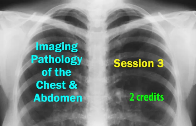
Chest radiography continues to be the most frequently ordered imaging exam due to its accessibility, low cost, low radiation dose, and high diagnostic value. This webinar explores foundational image assessment strategies used by radiologists to analyze chest radiographs, as well as the crucial contributions technologists make to ensure diagnostic-quality images. Participants will learn how to recognize radiographic patterns and anatomical landmarks that aid in the localization of pathology.
Concepts such as the Silhouette Sign, hilar overlay, cardiac curves, and chest consolidations will be reviewed, along with techniques for evaluating image quality and patient positioning. The session emphasizes the technologist's role in optimizing exposure, respiration, and positioning—factors that directly influence the diagnostic potential of each radiograph.
Agenda:
- Principles of Chest Image Assessment
- Five Radiographic Densities and Their Applications
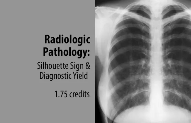
- The Silhouette Sign and Related Radiographic Concepts
- Types and Indicators of Chest Consolidation
- Cardio-Thoracic Ratio and Radiographic Analysis
- Identification of Cardiac Curves and Anatomical Landmarks
- Evaluation of Chest Lines, Stripes, and Other Key Structures
- Structured Approaches to Reviewing Chest Radiographs
- Technologist Strategies for Enhancing Diagnostic Image Value
- Positioning, Respiration, and Exposure Techniques
- Indicators of Image Rotation and Quality Variations
How it Works:
- The On-Demand CE activity that you purchased will be located in your “My Account” section once you log into the MTMI Website.
- You have three attempts to pass each quiz.
- You must earn a score of 75% or higher.
- Credit is recorded the day you submit and pass the quiz, and is determined using Central time.
- You have 30 days to complete and pass the quiz.
- Once passed, access your MTMI “My Account” to print your “Certificate of Completion.”
- This video expires 1 year after purchase date.
Educational Objectives
At the conclusion of this program, participants should be able to:
- Describe the five radiographic densities and their relevance in image assessment
- Differentiate the right and left hemidiaphragms on a lateral chest radiograph
- Recognize abnormal chest radiographs using the Silhouette Sign concept
- Identify the general location of chest consolidations using the Silhouette Sign and hilar overlay techniques
- Distinguish between the four classes of chest consolidations
- Diagram the pulmonary acinus to explain the process of fluid accumulation and aeration in lung tissues
- Calculate the cardio-thoracic ratio and explain its significance
- Label the left and right cardiac curves seen on chest radiographs
- Identify key anatomical structures commonly seen on chest radiographs
- Explain the technologist’s role in maximizing image quality and the consequences of suboptimal imaging
Program Faculty
Meet your presenter(s)

Randy Griswold
MPA, RT(R)
Randy has been in the medical imaging profession for over 40 years as an educator, sales and marketing professional and consultant. Currently he is a contributing author and medical imaging consultant. Prior to that, he was the Director of Sales and Marketing for a Midwest distributor of digital medical imaging products. His collective experiences as an educator include being the Program Director for Bellin College, School of Radiologic Sciences and their BSRS program. He was instrumental in transforming their two-year certificate program to a four-year, accredited BSRS degree program.
Randy has 23 years of experience in radiology capital equipment sales, service and support including digital imaging, and has completed his graduate work in Public Service Administration with an emphasis in Health Care Administration and Medical Imaging Marketing. He is a past president of the Wisconsin Association of Educators in Radiologic Technology (WAERT) and the Wisconsin Society of Radiologic Technologists (WSRT) as well as being a Fellow. His passion for teaching is focused on helping technologists understand the importance of obtaining good quality images for diagnosis, in a fashion that uses the skills and techniques of radiologist interpretation.
Credits
Accredited training programs
ASRT Category A
This program provides 2 hour(s) of Category A continuing education credit for radiologic technologists approved by ASRT and recognized by the ARRT® and various licensure states. Category A credit is also recognized for CE credit in Canada. You must attend the entire program to receive your certificate of completion.