

Trauma Radiography: Imaging Patients in Crisis
About this Program
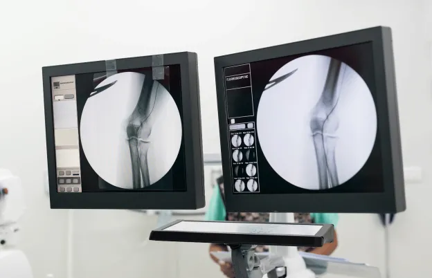
Recognition of the pathological processes present in your images is key to ensuring a successful imaging exam for trauma radiography. This presentation will introduce the methods and techniques to overcome non-routine challenges in diagnostic radiology. A major focus will be the inter-relationship of the various imaging modalities and how they clinically complement each other with imaging trauma patients. Select radiographic positions will be discussed as to clinical expectations in order to maximize diagnostic yield in an ALARA fashion and to give the participant a radiologist’s perspective as to the challenges in image interpretation. The speaker will be utilizing a case-based approach in the delivery of these concepts.
Educational Objectives
At the completion of this program, attendees will be able to:
- Outline the critical parameters of acceptable images in non-routine settings
- Suggest methods and techniques to overcome non-routine imaging challenges
- Analyze medical images for essential information in non-routine situations
- Discuss the role of SID and centering when imaging patients in trauma cases
- Adapt to unique situations when radiographing patients in non-routine settings
- Suggest modifications to routine positions to improve clinical utility in trauma and mobile radiography situations
- Explain the concept of silhouette sign and its clinical applications
- Discuss the role of fat as a radiographic density in radiographic image interpretation
- Identify the five (5) basic radiographic densities on all radiographic images
- Analyze acceptable image quality using clinical image identifiers and exposure index values
- Appreciate your role as a medical imaging professional in producing optimal images and as a patient advocate
- Discuss technological designs as they apply to imaging in trauma situations
Schedule
What this course will cover
Thinking Like a Radiologist?
|
Trauma Considerations
Chest Analysis in Trauma Situation
|
Lunch |
Image Analysis in Trauma Setting and Radiologist Expectations
|
Trauma Positioning for Body Parts with Clinical Image Expectations
Exposure Index (EI) and Deviation Index (DI)
|
Audience
Who should attend?
- Radiologic Technologists
- Medical Imaging Specialists
- Educators
- Vendor Personnel
Program Faculty
Meet your presenter(s)

Randy Griswold
MPA, RT(R)
Randy has been in the medical imaging profession for over 40 years as an educator, sales and marketing professional and consultant. Currently he is a contributing author and medical imaging consultant. Prior to that, he was the Director of Sales and Marketing for a Midwest distributor of digital medical imaging products. His collective experiences as an educator include being the Program Director for Bellin College, School of Radiologic Sciences and their BSRS program. He was instrumental in transforming their two-year certificate program to a four-year, accredited BSRS degree program.
Randy has 23 years of experience in radiology capital equipment sales, service and support including digital imaging, and has completed his graduate work in Public Service Administration with an emphasis in Health Care Administration and Medical Imaging Marketing. He is a past president of the Wisconsin Association of Educators in Radiologic Technology (WAERT) and the Wisconsin Society of Radiologic Technologists (WSRT) as well as being a Fellow. His passion for teaching is focused on helping technologists understand the importance of obtaining good quality images for diagnosis, in a fashion that uses the skills and techniques of radiologist interpretation.
Credits
Accredited training programs
ASRT Category A
This program provides 8 hour(s) of Category A continuing education credit for radiologic technologists approved by ASRT and recognized by the ARRT® and various licensure states. Category A credit is also recognized for CE credit in Canada. You must attend the entire program to receive your certificate of completion.
Tuition

| Audience | Price | Early Price | Member Price | Member Early Price |
|---|---|---|---|---|
| Technologist | $149.00 | $143.00 | $135.00 | $129.00 |
Early Pricing Guidelines
Qualifying 'Early' registrations must be made at least 14 days in advance for the program.
Cancellation Policy
Seminars/Webinars 8 hours of credit or more
Refunds, minus a $30 processing fee, will be granted for cancellations received at least 3 days prior to the program. Cancellations received within 3 days of the program will receive a credit toward a future MTMI program, minus the $30 processing fee. No refunds will be made after the program starts. MTMI reserves the right to cancel any scheduled program because of low advance registration or other reasons. MTMI’s liability is limited to a refund of any program tuition paid. MTMI recommends that attendees use refundable airline tickets. In case of cancellation of a program for any reason, MTMI is not responsible for travel costs incurred by attendees including non-refundable airline tickets. When offered, WEBINAR ATTENDEES that cannot log in due to unsolvable technical issues beyond their control will be eligible for a full refund.